Description
Technology
Left, Top
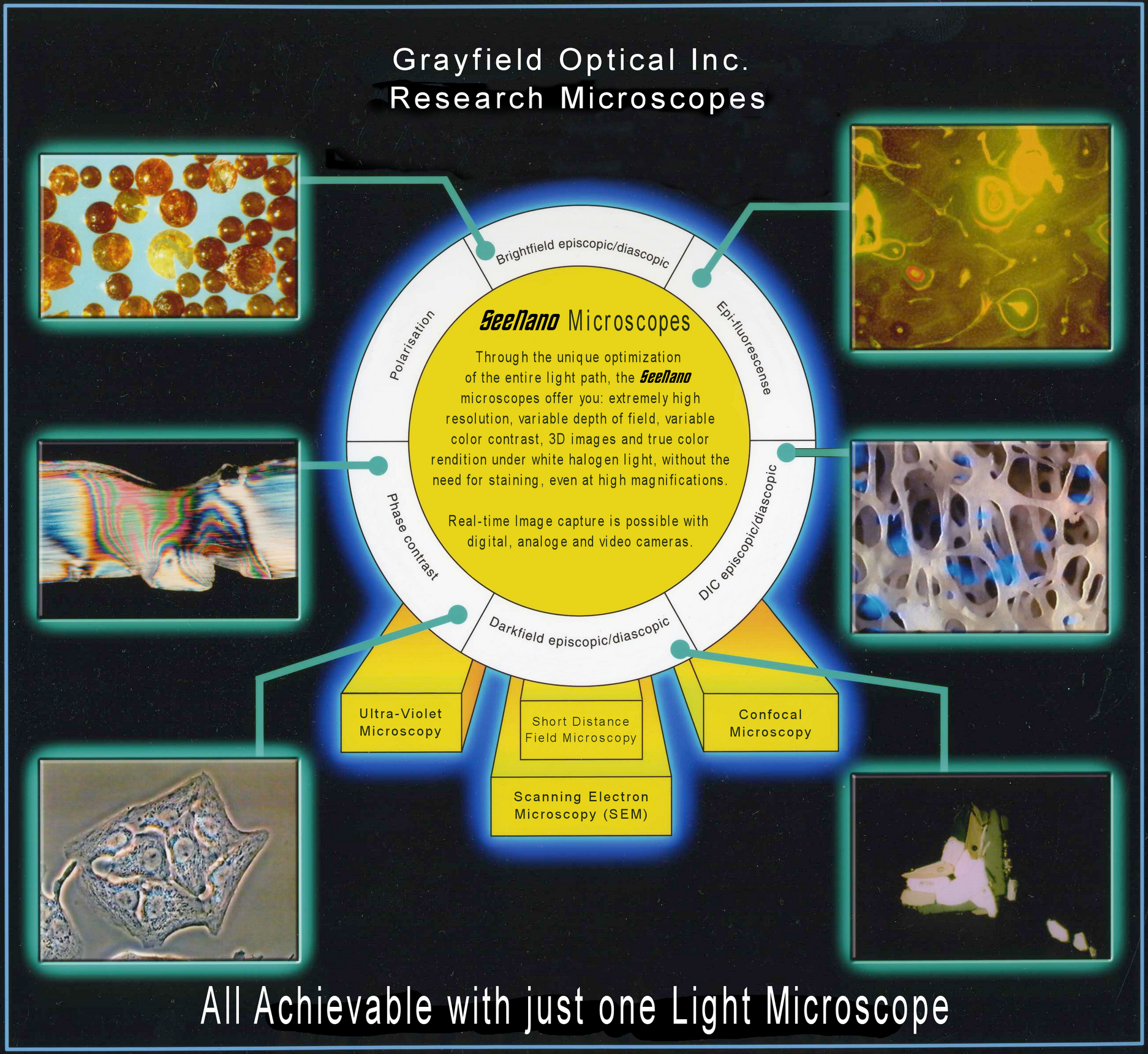
Tiny pellets of methyl methacrylate MMA (Plexiglas®). This batch has been polluted by a soot explosion and can no longer be used to manufacture transparent products.
Left, Center
Thin section of a plastic tube press connection. The predetermined break point is clearly visible in polarized light.
Left, Bottom
Living heart muscle seen in vitro under phase contrast with enhanced depth of field.
Right, Top
Cross section of a bone from a racing horse seen using Epi-fluorescense.
Right, Center
Structures of a 3.5 mm thick cross section of a bone. Extreme depth of field allows entire structure to be seen clearly.
Right, Bottom
Crystals in water under polarised light as imaged using an extended colour spectrum possible with the SeeNano.
"Those who say it cannot be done should not get in the way of the person doing it" - Chinese proverb.
Introduction
Ever since the German physicist Ernst Abbe determined that light microscopes cannot resolve objects smaller than half the wavelength of visible light (about 250nm), this has been seen by scientists as the absolute resolution limit of light optical microscopes.
The current Grayfield Lens System (GLS) is the result of over 40 years research and development. By closely examining and optimising every part of the optical pathway and closely reexaming the laws of optics, putting every aspect of those laws into question, a unique way of designing optical systems was discovered, where the limits normally associated with optical resolution simply do not apply.
We recognise that many of the statements, images and videos shown on this site regarding the optical capabilities of our microscopes will appear incredible to those who have studied the existing laws of optical physics. Yet these capabilities are real and based on solid optical designs which work extremely reliably, as anyone who has uses our microscopes will discover.
True scientists should remain open for new scientific developments and we ask you to reserve judgement while you view the numerous images, videos and data and ask yourself if the capabilities of our optical systems, as presented on this website, would be useful to your research work. Anyone visiting our labs can see for themselves what can really be achieved with this technology.
On this website, we will present a considerable amount of proof for you to judge for yourself what is possible with this technology.
We thank-you for your interest.
Light Microscopy with SeeNano-Technology
No Staining, No Oil immersion, Extended Depth of Field, Full Color Contrast, Natural Colors, Full Contour Sharpness
The SeeNano technology introduces a new method of providing a much higher depth of field and contrast, where the natural colors (white light source) and contour sharpness remain clearly decernable even with ever increasing magnification.
This allows a quality of observation that can meet the highest and most demanding requirements. Only the real time analysis (In-Situ) of living microorgnisms (In-Vivo, in-Vitro) and materials can provide a truely realistic insight. This means that samples can be observed optically, without time consuming preparation (staining, etc.), under normal room temperatures.
These capabilities are controlled, thanks to a sophisticated mechanism, using just one additional control. In contrast to current light microscopes, the SeeNano microscopes fully maintain parallax free images.
The angle of view, by which the object is imaged, can be modified between vertical (90°) and diagonal (45°). No staining or complicated sample preparation is required. All our objectives are designed for dry use. No messy oil immersion is required or desired to obtain the high resolutions (<100nm under reflected light) available. The variable depth of field allows you to see uneven surfaces in sharp contrast or alternatively to see individual layers of living tissues without the higher or lower layers interfering with the image quality. Even at high magnifications, we can still maintain a greater than normal working distance, typically 1-3mm, it is rarely necessary for the objective lens to actually touch the object.
New Standards for Light Microscopy
Phase Contrast without over-exposure!
The phase contrast method is used for all thin-layered structures including fibres and textiles. With conventional methods, focussing is limited by overlapping layers and the structures also have the same fractal index causing the structures to become blurred. The SeeNano technology removes these limitations and provides clear and sharp contours.
Standardized objectives for color contrast, transmitted light, darkfield and polarisation
The SeeNano-technology makes it possible to use the same objectives for phase contrast, transmitted light, darkfield, grayfield and polarisation. Until now, polarisation required not only a separate microscope, but also special polorisation objectives. Without such objectives the phase rings would be seen as distortions when using transmitted light.
Grayfield Contrast Method
An entirely new method in optical microscopy is the grayfield contrast method. This method allows you to see detailed structures that are otherwise not even visible with conventional phase contrast microscopes. For example, the grayfield method allows you to observe the in-vitro decomposition processes of blood. During this transitional phase new viruses and structures arise, which tend to decay and could previously not be made visible due to the lack of suitable microscope techniques.
Confocal method:
This very interesting feature makes it possible to switch very quickly between vertical and an angle (from 45° to horizontal).
This enables the following:
- Following the movment of viruses
- Combining the momentum of viruses with magnetic fields ("Freezing" of living procedures (1/1000 sec) for a still image observation
- Intruding into translucent surfaces with complete analysis within 2 - 3 minutes (digital, analog or in "slow motion")
- In-Vivo-Focussing of human arteries with "magnetic freezing" to allow for immediate analysis of all cells in (flowing) blood to allow analyisation of the substances contained (duration ca. 1-2 seconds).
- Is comparable to an endoscope with a magnetic field "Blocker" on the probe
Renewal of product lines at Grayfield Optical, Inc.
We previously offered the "Ergonom" range of microscopes with pricing often beyond the range of many doctors and clinics. Those microscopes are now no longer available as the inventor, Kurt Olbrich, retired and later died in January 2022. We wish to thank him for all he has done in the past including his development work on the series of microscopes we have been offering.
Grayfield Optical are now working with his engineer, that has been working with this microscope technology for decades, is considerably younger and fully up-to-date with the latest microscope technologies including the latest digital measurment systems. Our nano-positioning system was developed by this engineer, for example.
We are now preparing to release an entirely new series of microscopes, under the SeeNano name, which enables us to provide you with microscopes that have all the capabilities of the previous series while adding fluorescence capabilities and much more realistic pricing. This new series should be available from Q3, 2022 and we ask you to be patient while we complete work on a new series of microscopes.