References
As Kurt Olbrich has a background in industry, most of the work done was on contract with companies in the automotive, plastics, metal and electronic industries with only very limited research in the field of medicine. Most of the work done was on contract to industry and state research agencies and subject to non-disclosure agreements.
As Kurt Olbrich did not speak English, most of his work was done for German speaking organizations and the references are therefore often in German.
A major breakthrough was made with the development of the Ergonom 400 microscopes during 1976 (in fact he built several different versions of the Ergonom 400 as shown in the history).
These microscopes made use of the Grayfield method, a new technology developed by Kurt Olbrich, which enables unstained samples to be seen not as outlines, but as clear sharp images with full color contrast even at high resolutions. The image detail obtainable with this method allows the viewing of live biological samples in a quality never seen before. Although these older microscopes were difficult and complicated to use, the capabilities of these microscopes, along with the ingeniuty of Kurt Olbrich in solving difficult problems, led to his institute gaining the reputation of being able to solve problems, nobody else could.
Many scientific reseachers in Germany often say: "if your research has come to a dead end - go to Olbrich!".
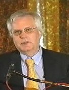
Dr. Gary L. Greenberg, Ph. D.
Kurt Olbrich has sold over 20 of his "Olbrich 4000" microscopes (based on the Ergonom 400) to various research organisations throughout the world. After writing this reference, Dr. Gary Greenberg, PhD arranged for the purchase of such a microscope for use at the University College London, Dept. of Anatomy and Developmental Biology. They have reported that the greater resolution and magnification possible allows them to greatly reduce research time.
A video including their research work can be seen in the Video section.
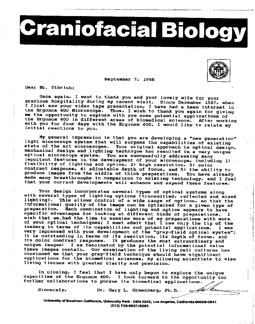
Craniofacial Biology
September 7, 1988
Dear Mr. Olbrich,
Once again, I want to thank you and your lovely wife for your gracious hospitality during my recent visit. Since December 1987, when I first saw your video tape presentation, I have had a keen interest in the Ergonom 400 Microscope. Thus, I wish to thank you again for giving me the opportunity to explore with you some potential applications of the Ergonom 400 in different areas of biomedical science. After working with you for four days with the Ergonom 400, I would like to relate my initial reactions to you.
My general impression is that you are developing a "new generation" light microscope system that will surpass the capabilities of existing state of the art microscopes. Your original approach to optical design, mechanical design and lighting technique has resulted in a very unique optical microscope system. You are successfully addressing many important features in the development of your microscope, including 1) flexibility of lighting and optics, 2) high resolution, 3) color contrast response, 4) adjustable depth of focus, and 5) the ability to produce images from the middle of thick preparations. You have already made many breakthroughs in comparison to existing technology, and I feel that your current developments will enhance and expand these features.
In closing, I feel that I have only begun to explore the unique capacities of the Ergonom 400. I look forward to the opportunity for further collaborations to pursue its biomedical applications.
Sincerely, Dr. Gary L. Greenberg* Ph.D.
University of Southern California, University Park - DEN 4242, Los Angeles, California 90080-0641
Prof. Gerd Binning (Nobel prize for Physics in 1986 for the scanning tunneling microscope) made extensive use of the Ergonom 400 in the early 1990s where he said:
(Click on image on left to see the original German letter)

Dr. Dr. Kurt S. Zänker
Prof. for Immunology
Witten/Herdecke University
Germany
Adjunct Professor
McGill-University Montreal
Canada
Kurt Olbrich was also involved in medical research work in colaboration with Bernhard Muschlien as well as numerous researchers at the University of Heidelberg, Germany.
In 1993, the Erlangen and Witten/Herdecke Universities (Germany) tested the Ergonom 400 microscope in the field of Immunology. Dr. U. G. Randoll (Erlangen) and Dr. Dr. Kurt S. Zänder, Professor for Immunology (Witten/Herdecke) conducted the tests and wrote a short expertise of their findings where for example Dr. Dr. Prof. Zänder commented that
"Both the resolution and the depth of field are significantly better than existing light microscopes when observing cell preparations (cell cultures)."
SHORT EXPERTISE ON THE ERGONOM 400
In cooperation with Mr. K. Olbrich, we were able to gain experience in the biological application of his new light optical microscope system. The preliminary results we have achieved so far, in cooperation with the University of Erlangen (Dr. U. G. Randoll), can be summarised as follows:
1. Both the resolution and the depth of field are significantly better than existing light microscopes when observing cell preparations (cell cultures).
2. A further advantage, when compared with existing light microscopes, is that native, unstained preparations, like smear tests or biopsies (living cell samples from patients), could be directly examined in a new previously unseen dimension, where for the first time, even the non-balanced thermodynamic expectations of modern cell biological diagnostics could be fulfilled.
3. The Ergonom 400 allows the immediate observation of unprepared preparations (e.g. by staining, fluorescence, or sputter), it however does not exclude such methods. The spatial resolution lies in the range of a mid-range Scanning Electron Microscope.
EVALUATION
The microscope and ultra-structure tomography system (living preparations can be focussed through layer by layer where each layer is shown sharply), that has been developed by Kurt Olbrich, is an innovative contribution to the development of imaging systems. Mr. Olbrich has made this development in an admirably persistent and purposeful way. This development might be unique in the area of microscopy at this time.
The Ergonom technology closes the gap, for the various scientific areas of research and development, between the top light microscopes and scanning electron microscopy. It is to be expected that, through the implementation of the various Ergonom range (of microscopes) in cell biology, virology and in cancer research, a considerable gain in knowledge can be achieved in the shortest time.
Through the advantages of native observation and the cellular tomography, a significant contribution has been made in order to reduce animal testing in the areas of pharmacology and environmental toxicology.
It must also be emphasised that the work with such microscopes will lead to a considerable requirement for research in regard to the accompanying results.
Witten 5th Feb. 1993
Witten/Herdecke University
Adjunct Professor, McGill-University Montreal, Canada
(Translation of the original German text. Click on image to see the original)
Institute for Immunology
Witten/Herdecke University
Prof. Dr. K. S. Zänker
Stockumer Str. 10
58453 Witten
Germany
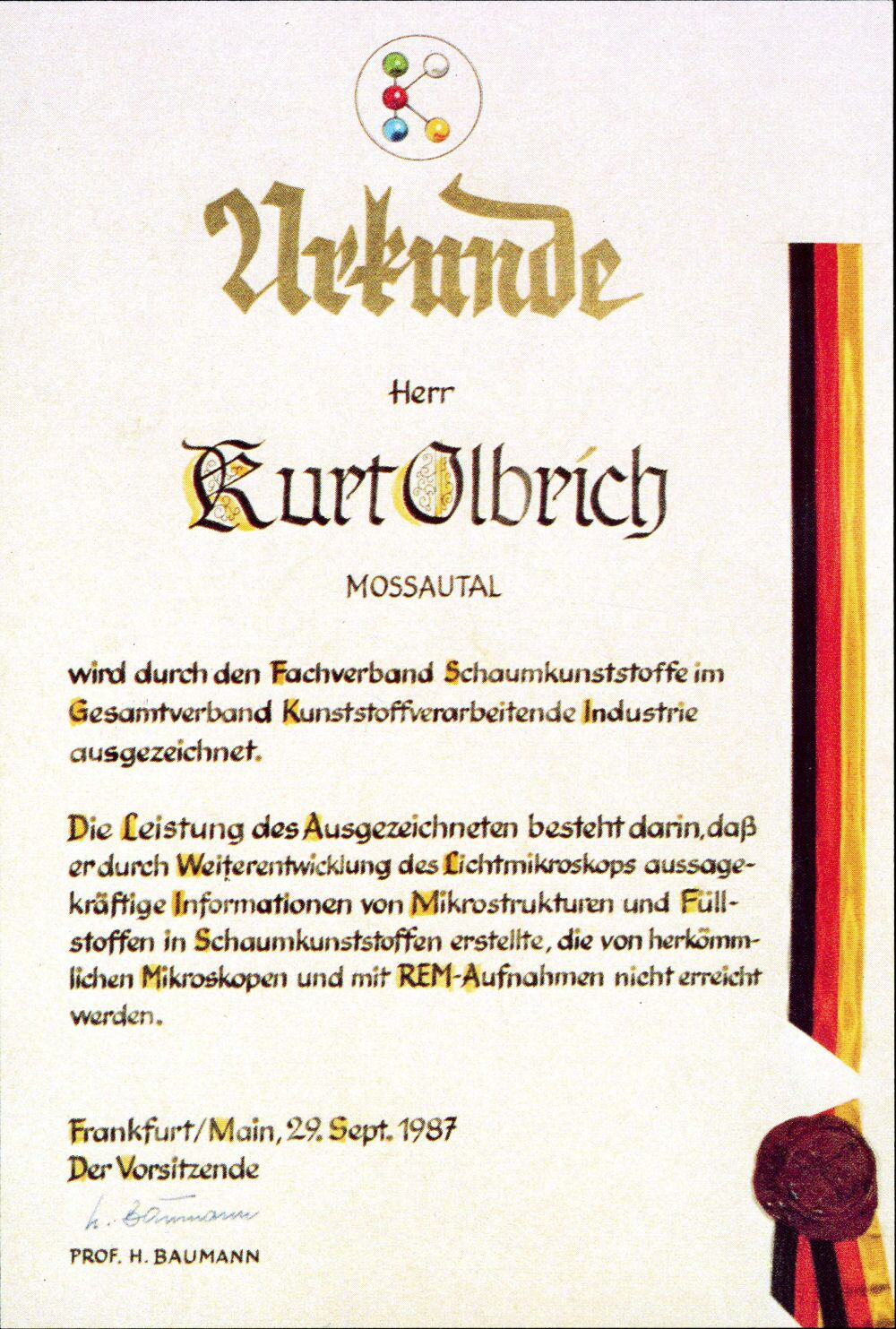
Certificate of the German Plastics Association
Until recently (he has now retired from that job), Kurt Olbrich was a German state authorized consultant for analysis of plastics.
On September 29th, 1987 he received a certificate honoring his work from the German plastics association.
Here is a translation of that certificate (click on image to see the original):
Certificate
Mr.
Kurt Olbrich
Mossautal
(The town where he resides)
is being honored by the foam plastics section of the Plastics Processing Industry Association.
The achievement of the bearer of this certificate consists in the fact that through further development of the optical microscope, he provided meaningful information from microstructures and fillers in foamed plastics, which was not achievable with conventional microscopes and with SEM photographs.
Frankfurt/Main, 29th September 1987
The Director
Prof. H. Baumann