Medical
Examples > SeeNano Pro
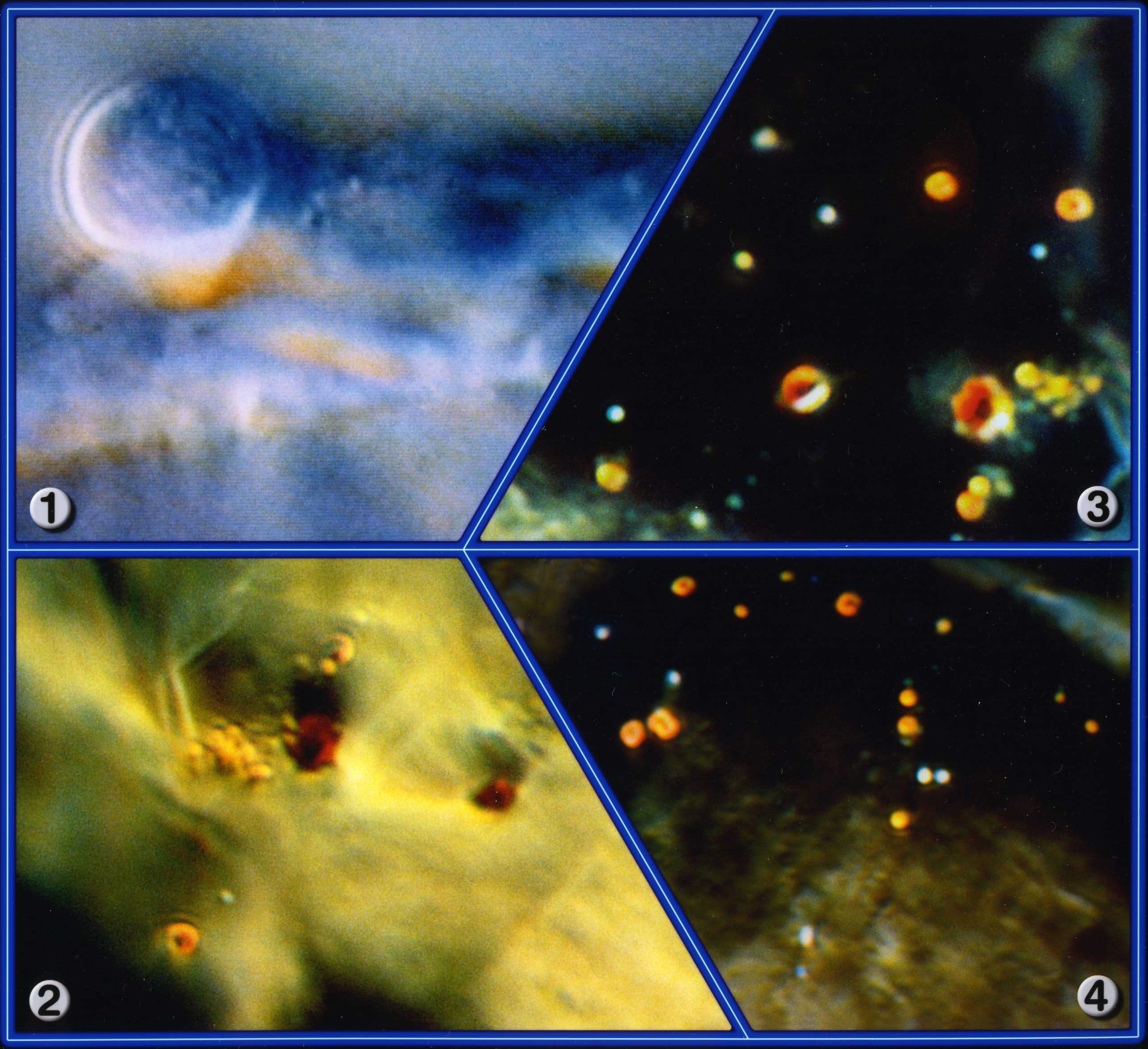
All Images: Magnification: 9000x
- Image 1-4: Cell tissue images from an Aids pure culture at the different stages of development.
The images made of the AIDS culture is interesting as scanning electron microscope (SEM) images shows them to be much smaller than in our images. After further observations, we found that the process of preparing such cultures to be observed with SEM makes them shrivel up and die. What you see under a scanning electron microscope is therefore not representative of their natural size and appearance. We were able to show that the AIDS causing pathogens, the orange colored specs, grow and in the fully grown form open up like a poppy flower (image 3).
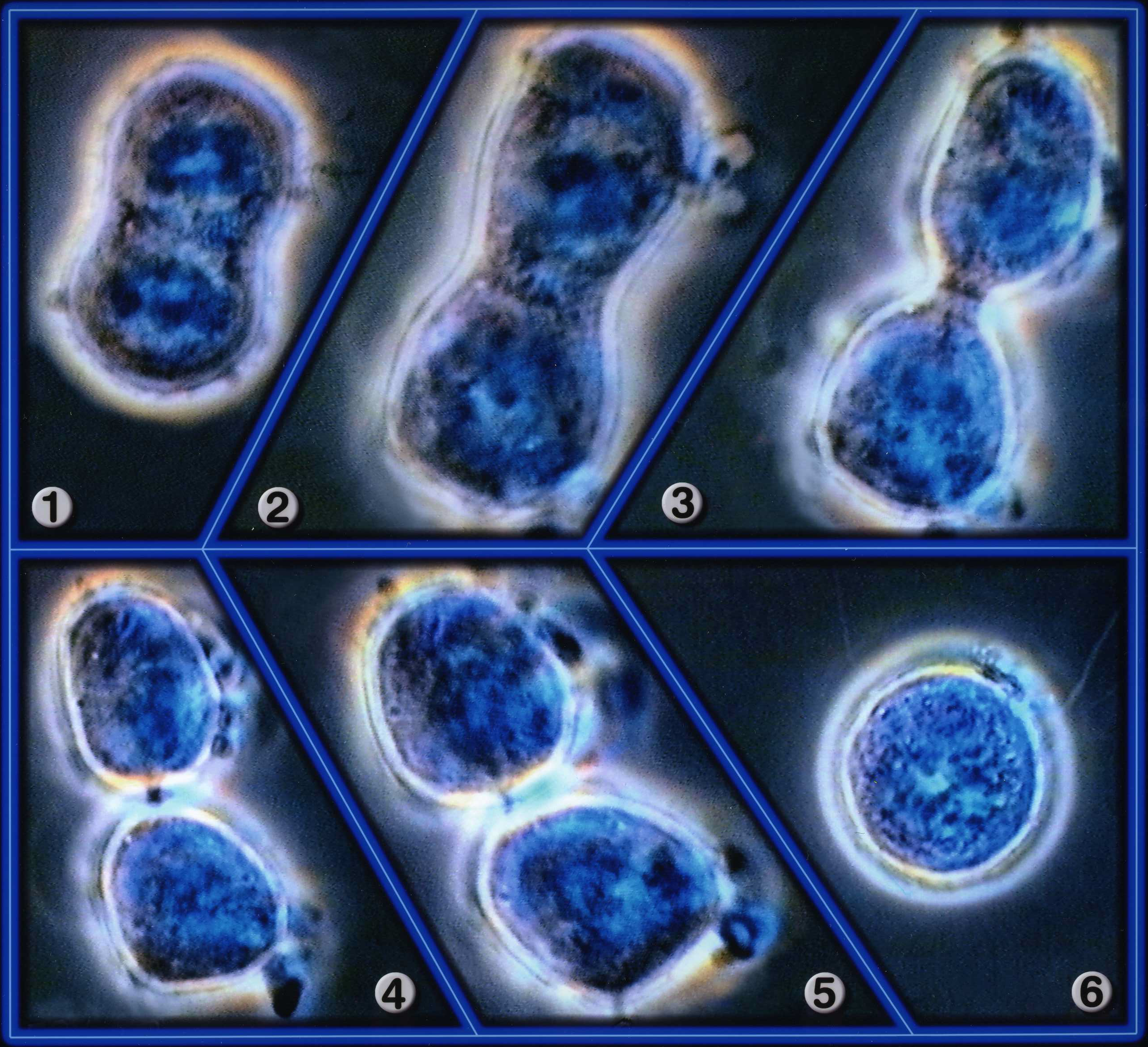
All Images: Magnification: 3570x
- Image 1: A cell that is undergoing division. Chromosome segregation has already occurred.
- Image 2: The constriction stage has been initiated.
- Image 3: Constriction between mother and daughter cells, each with half set of chromosomes.
- Image 4: Membrane organization completed between both cells.
- Image 5: Mother and daughter cells separate, mitotic process is complete.
- Image 6: Each cell is self-sufficient and can initiate cell division again.

Image Magnification: 14x
- Small screw made of PM for surgical use. The yellow arrow shows a defective area, which cannot be kept sterile and may become contaminated with bacteria.
Please note, we have many more medical images on our files and we will be adding more images, and better descriptions, shortly.
We also recommend viewing the online films
Click on the following images to see individual video clips taken from our films.
Cell Division
White blood cells
Bone structure in 3D
Cancer Tumor