SeeNano versus Light Optical Microscopes - Grayfield Optical Inc - High Resolution Optical Microscopes

Go to content

SeeNano versus Light Optical Microscopes

Comparison

The following table compares the capabilities of the SeeNano Microscope with conventional top quality light microscopes:

Reflected Light - Microscopes

Conventional Light Microscope
"State of the Art"
Unchanged since the 19th century
SeeNano Microscope
Grayfield-Technology
The new standard for the 21st century


Transmitted Light - Microscopes

Conventional Light Microscope
Light reflections cause over exposure (hazy picture). Reduction of over exposure by adjustment of light field aperture which unfortunately also reduces contour sharpness.

Depth of Field:

Marginal, declines with magnification

Resolution (max):

Practically    : 500nm
Theoretically: 250nm

Natural Colors:

Not possible at high resolutions

Sample warmth levels:

Up to 60 °C above room temperature

Use of Living Materials:

Strictly limited due to high warmth levels. Living cells typically survive for only a few minutes.

SeeNano Microscope
No light reflection and no over exposure.
This is the basis for brilliant pictures. Iris aperture used instead of light field aperture. Extremely high contour sharpness.

Depth of Field:

Extremely high, infinitely variable, independent of magnification

Resolution (max):

Practically    : 100nm
Theoretically:   10nm

Natural Colors:

Unlimited up to the most extreme resolutions

Sample warmth levels:

Max. 5 °C (typ. 2 °C) above room temperature

Use of Living Materials:

Limited only by natural life span of cells.
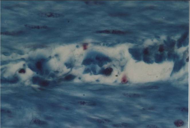
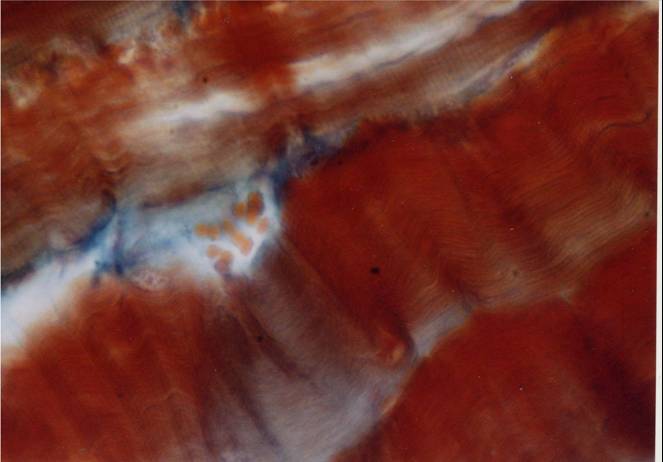
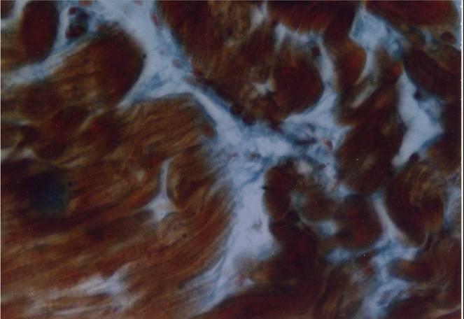
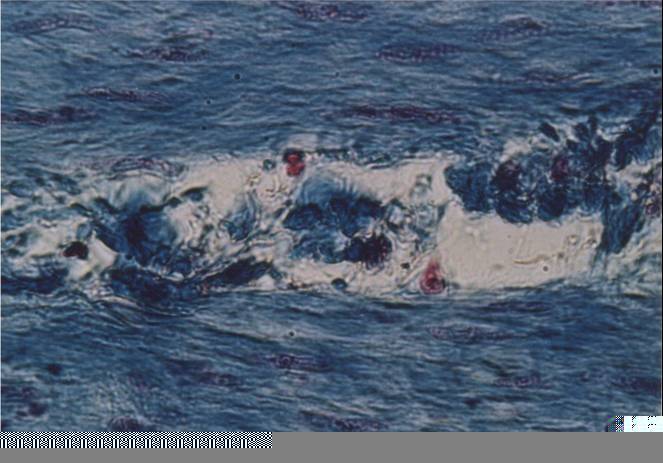
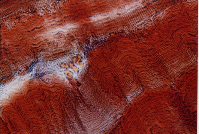
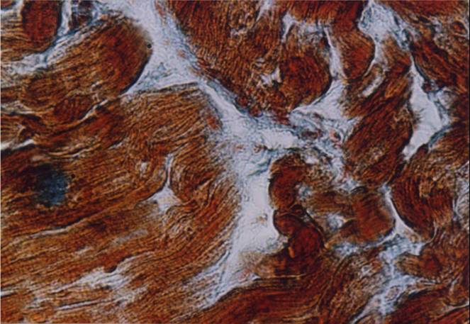
Use of chambers make long-term observations possible. e.g. 2-3 weeks on nerve cells up to several months on the pathogens of the legionnaire´s disease


Stained Slides


The Grayfield microscopes do not require any form of staining or time consuming preparation. This allows the specimens to be seen in their natural state in natural colors and in high detail.
However, in order to compare the SeeNano with conventional light microscopes, the following images were made with stained slides under a top quality conventional light microscope and the SeeNano Pro. Despite the staining, it is obvious that the SeeNano can reveal considerably more details than the conventional light microscope.
Conventional Light Microscope
SeeNano Microscope
© Copyright 1976-2023: Grayfield Optical, Inc.
© 1976-2017: Grayfield Optical, Inc.
Back to content