SeeNano versus Scanning Electron Microscope
Comparison
Scanning Electron Microscope
A scanning electron microscope can provide a considerable amount of depth of field (greater than that of the SeeNano system) and very high quality black and white images. The resolution capabilities of the SeeNano microscopes reach into the realms of mid-range SEM while offering a number of advantages:
- No sputter coating or staining required
- No vacuum required, viewed in normal room conditions
- Sample does not need to be cut to a small size to fit in a chamber
- Non destructive for living organisms
- SEM images are made at 45° angle causing image distortion!
- SeeNano images can be made at any angle between 45-90°
- Measurable in X, Y and Z axis
- Natural colors and grayfield contrast provide more image information
- Confocal imaging allows viewing below the surface of tissue samples
Scanning Electron Microscope

SEM image of computer chip as photographed at 45° angle in B&W
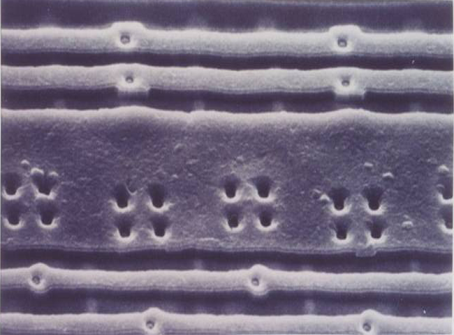
SEM Image corrected (stretched) to simulate 90° angle
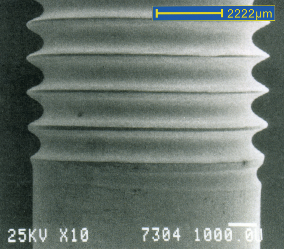
Screw used in automotive applications. SEM
SeeNano Microscope
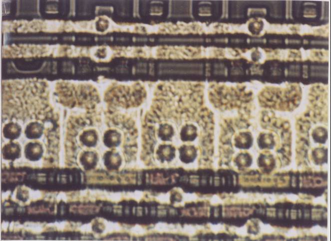
Photographed at 90° angle with DOF in color
Images precisely in both X and Y axis at 90°
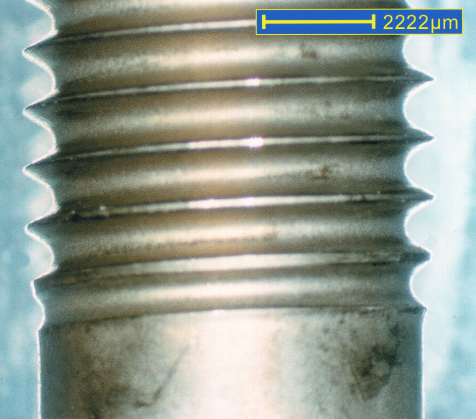
Screw used in automotive applications.